Lung cancer claims the lives of more than 150,000 people each year, with nearly a quarter of a million new cases being diagnosed every single year. That makes lung cancer the leading cause of cancer death for both men and women, more than breast, colon, and prostate cancers combined.
With lung cancer being so prevalent, early detection is key and anything that can be done to improve that process could save many lives each year. Advances in technology have now made it possible to use low-dose CT scans to help diagnose lung cancer at a very early stage while it’s still very treatable.
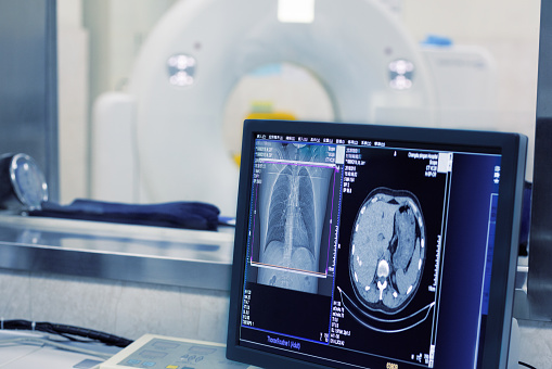
A computed tomography (CT) scan produces cross-sectional images using several X-ray measurements taken from various angles. The technology was introduced in the 1970s but has recently been used to detect lung cancer.
The patient lies on a table as the CT scanner takes several pictures while it rotates around them. A computer combines the pictures to give a more complete image which can show lung tumors more effectively than a routine chest X-ray. The technology can also show the shape, size, and position of tumors in addition to showing enlarged lymph nodes that could contain cancer that’s spread from the lungs.
Because lung cancer symptoms aren’t usually present until the advanced stages of the disease, screening for high-risk patients is especially important. Medicare even covers the cost of these screenings for patients aged 55 to 77 who have smoked at least one pack of cigarettes a day for 30 years and who still smoke or have only quit within the last 15 years. According to the American Lung Association, about nine million people fit the criteria, but many of them don’t know about the procedure.
Contact Salem Radiology today at 503-399-1262 to schedule an appointment.

